Mitochondria from a woman's leg were injected into her eyes
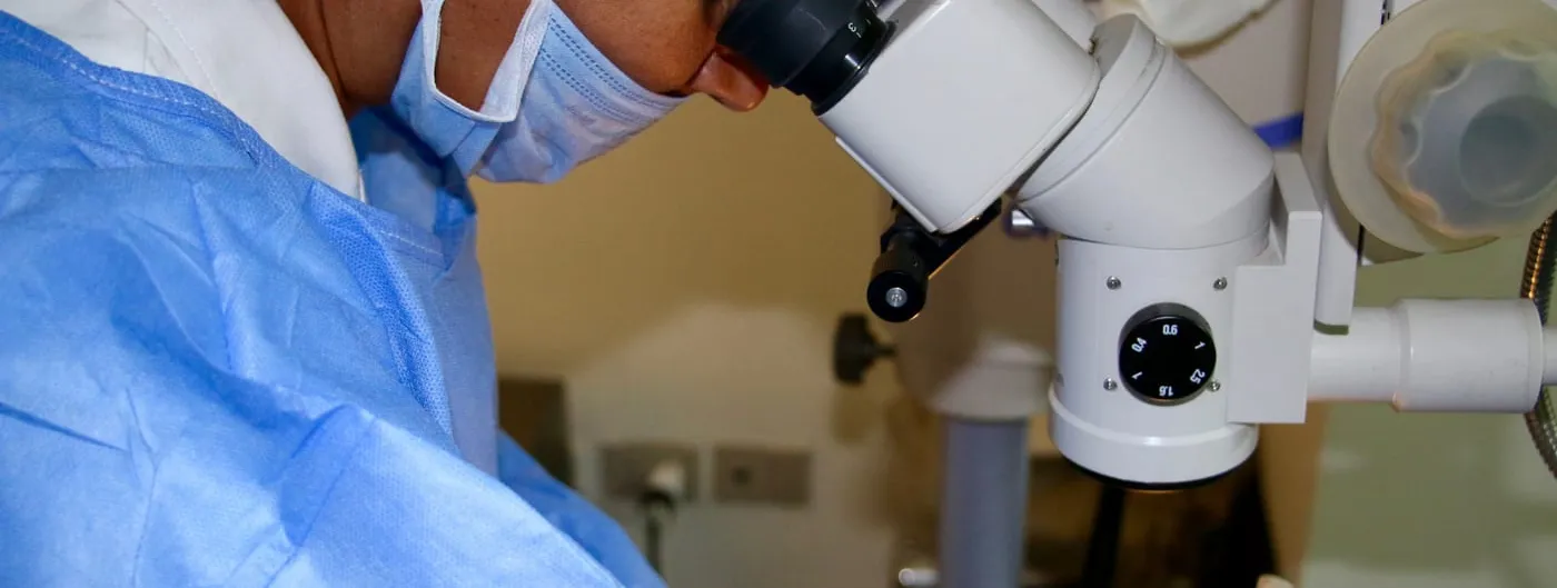

A team led by neuroscientist David Putrino at the Icahn School of Medicine at Mount Sinai took mitochondria from a thigh-muscle biopsy of a 26-year-old woman and injected them into the vitreous of each of her eyes, the two injections 24 hours apart. She had been left with almost no vision by bilateral optic neuropathy following a brain bleed, and the procedure was carried out under emergency expanded access about three months after the injury.
Before treatment, none of 45 measurements showed a normal pupil response to light. Within days of the injections, responses appeared: they persisted through day 11 in the left eye and day 39 in the right, then faded. Organised activity was detected in the visual cortex at 3, 44 and 45 days. Visual acuity did not change; she perceives light, not form. No ocular or systemic toxicity was observed.
The report is a preprint on Research Square and has not been peer reviewed. Its authors say plainly that a single uncontrolled case cannot establish causation. What it does establish is that freshly isolated autologous mitochondria can be placed in a human eye without apparent harm, which is the question a first case exists to answer.