Human organoids grow into more than 90% of a mouse cortex
Pasca lab / Stanford MedicinePress kit
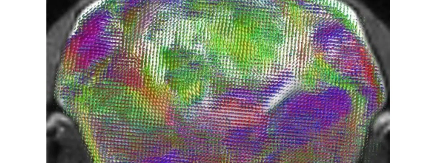
Sergiu Pasca’s laboratory at Stanford Medicine reported on 16 September 2026 a mouse whose cerebral cortex is mostly human tissue. The team bred apallial mice, genetically engineered so that almost none of their own neocortex and hippocampus forms, then grafted laboratory-grown human cortical organoids into the newborn animals. They call the procedure xenocortication.
Three months after surgery, human tissue accounted for more than 90% of cortical volume. Human and mouse neurons formed integrated circuits, and between three and six months after the graft the xenocortical mice performed like ordinary mice on behavioural tests; working-memory deficits seen in the cortex-depleted animals were reversed by the transplants. The human tissue matured further than organoids grown in a dish, and included von Economo neurons, a cell type found in large-brained mammals that had been observed only in post-mortem tissue and never generated in culture.
The model is built for disease. Depriving the animals of oxygen damaged the human tissue selectively, with immune-cell infiltration and stress markers of the kind seen after brain injury, and produced deficits resembling cerebral palsy. Because the grafted cells carry the donor’s genome, a patient’s mutations can be followed into circuits rather than cell cultures.
Pasca convened an Asilomar conference on the ethics of the work in November 2025, with ethicists, philosophers and legal scholars. The paper was published in Nature.