Twenty minutes of stimulation shifted gene activity in brain tissue
UT Southwestern Medical CenterPress kit
UT Southwestern Medical Center reported on 10 August 2026 a study in Nature that asks what electrical stimulation actually does to human brain tissue at the level of cells and genes. Bradley Lega at UT Southwestern and Genevieve Konopka, now chair of neurobiology at UCLA, worked from tissue removed during epilepsy surgery at UT Southwestern and Parkland Memorial Hospital and kept alive in culture for days afterwards, with Haley Moore as first author.
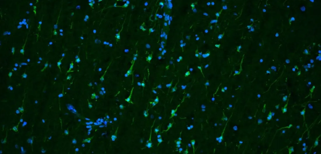
The group identified 19 neuronal assemblies — groups of cells that fire together — across tissue from 12 patients, and validated its observations against eight patients who had undergone deep brain stimulation. Twenty minutes of stimulation strengthened how those assemblies fired and increased their plasticity, their capacity to add or drop member cells. It also changed gene activity, in programmes governing neuronal activation, plasticity and synaptic communication, and not only in neurons: the supporting glial cells shifted too.
Deep brain stimulation has treated Parkinson’s disease and essential tremor for decades without a settled account of why it works, which has left stimulation protocols largely empirical. A mechanism at the level of gene expression is what a targeting rule would have to be built on.